Ecg Frontal Axis P Normal Range The P wave PR interval and PR segment ECG interpretation traditionally starts with an assessment of the P wave The P wave reflects atrial depolarization activation The PR interval is the distance between the
PR interval 120 200 milliseconds PR segment 50 120 milliseconds QRS complex 80 100 milliseconds ST segment 80 120 milliseconds T wave 160 NORMAL QRS axis Between 30 degrees and 90 degrees is normal There is much variation between individuals Even changing your position can change the axis of your
Ecg Frontal Axis P Normal Range
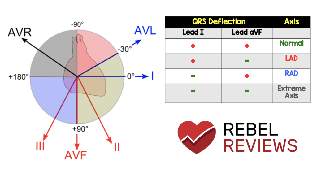
Ecg Frontal Axis P Normal Range
https://rebelem.com/wp-content/uploads/2019/03/ECG-Axis-1024x575.png
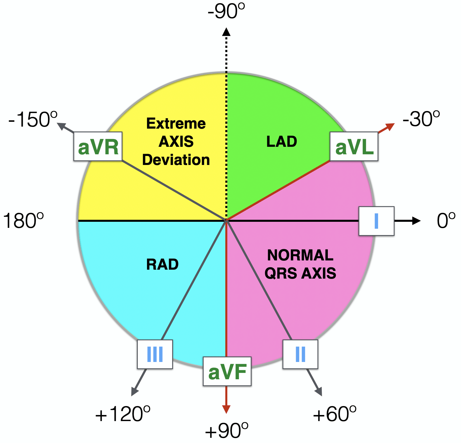
ECG Axis Interpretation LITFL ECG Library Basics
https://litfl.com/wp-content/uploads/2018/08/Haxaxial-ECG-AXIS-Reference-2021.png
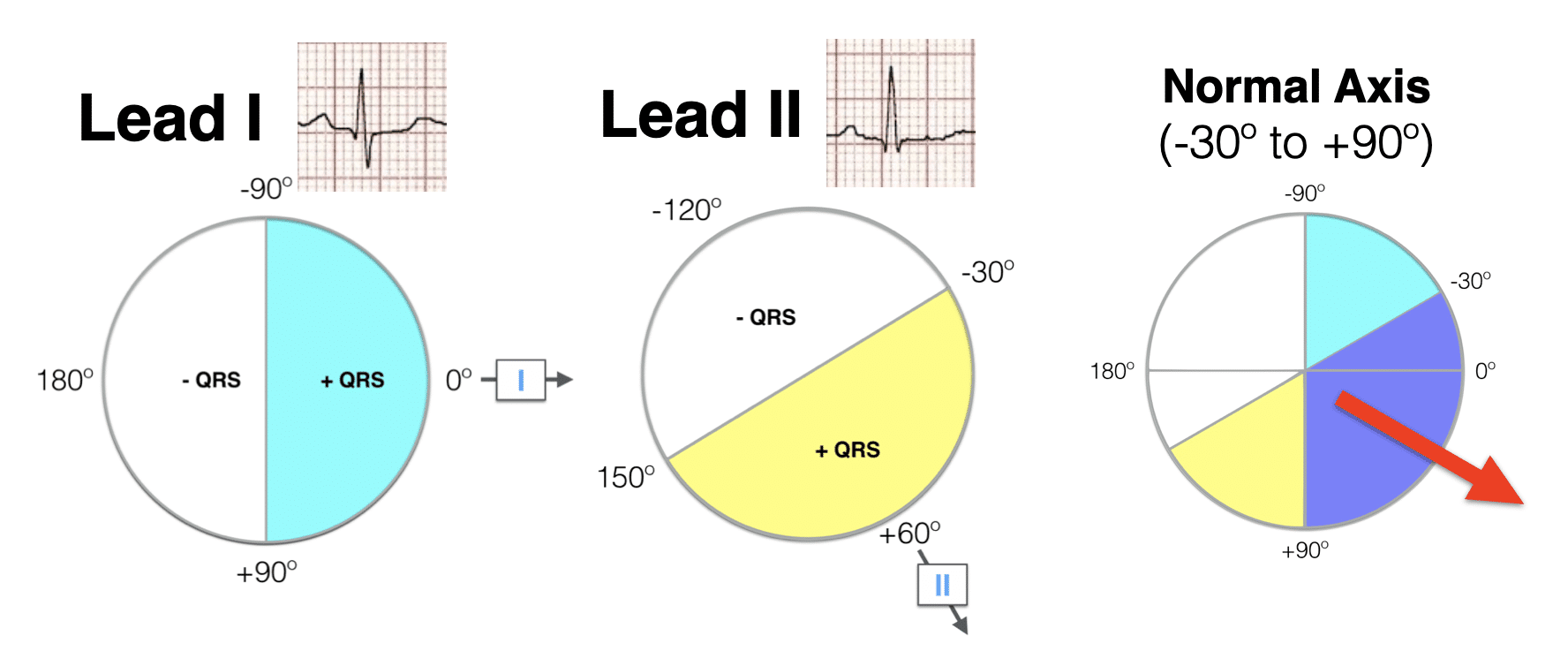
ECG Axis Interpretation LITFL ECG Library Basics
https://litfl.com/wp-content/uploads/2018/08/Lead-I-Lead-II-hexaxial-analysis-Normal-axis-2021.png
P wave always positive in lead II actually always positive in leads II III and aVF P wave duration should be Normal P wave axis is between 0 and 75 P waves should be upright in leads I and II inverted in aVR Duration 0 12 s
Each of the 4 quadrants represents 90 and an axis type eg 0 to 90 is a normal axis 90 to 180 is RAD 0 to 90 is LAD and 90 degrees to 180 degrees is an extreme axis Therefore if leads I and The P wave can have multiple measured parameters such as 1 duration 2 morphology 3 voltage 4 spatial axis and 5 area 8 P wave parameters PWPs can be combined to yield an index P wave
Download Ecg Frontal Axis P Normal Range
More picture related to Ecg Frontal Axis P Normal Range

EKG Interpretation Axis YouTube
https://i.ytimg.com/vi/grFic4Gb0zA/maxresdefault.jpg

Heart Axis Calculation
https://en.my-ekg.com/imag/normal-axis.png
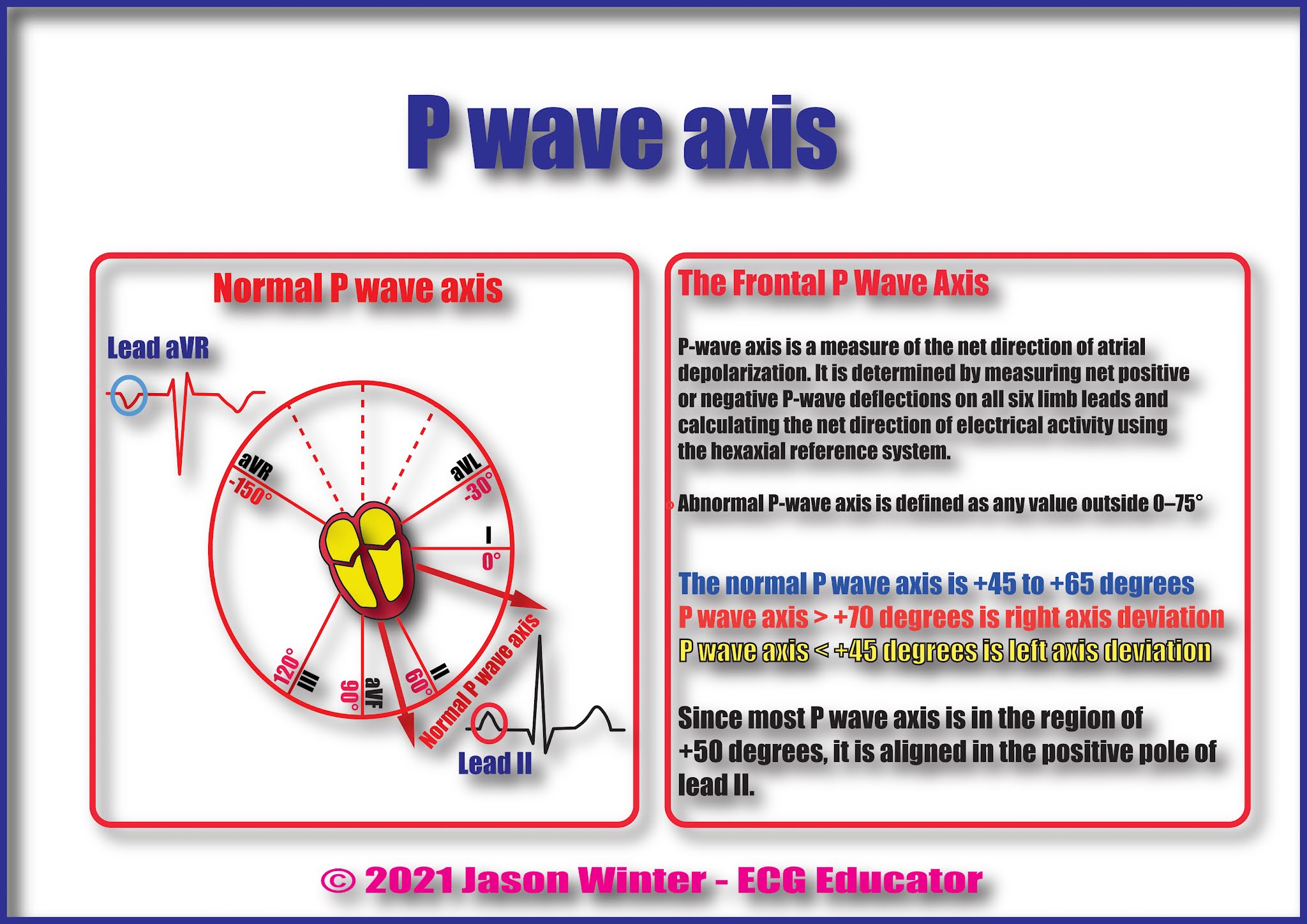
ECG Educator Blog P Axis On An ECG
https://1.bp.blogspot.com/-cl5Cfa-xXkw/YSeQuMXjWlI/AAAAAAAAD0M/UK9WEQUX9ikfkWVa7nW2adXU5hT84b0oACLcBGAsYHQ/s2048/P-wave%2Baxis%2Bblog.jpg
As evident from the figure the normal heart axis is between 30 and 90 If the axis is more positive than 90 it is referred to as right axis deviation If the axis is more negative than 30 it is referred to as left axis deviation The median frontal P axis is stable over the various age groups at about 60 for both sexes in the direction of lead II The median QRS axis remains at about 70 for
This large study provided normal values for heart rate P QRS and T frontal axis P and QRS overall duration PR and QT overall intervals and QTc corrected by Find the perpendiculars for each lead and chose an approximate QRS axis within the 30 range Occasionally each of the 6 frontal plane leads is small and or isoelectric The axis
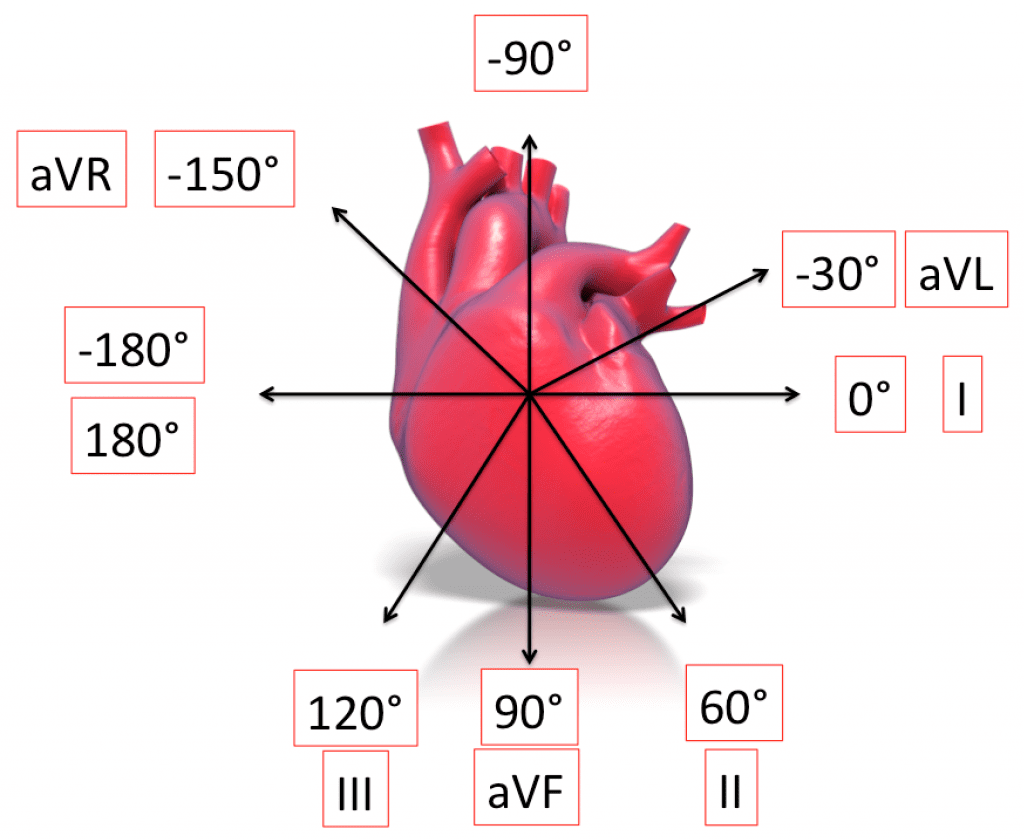
ECG Basics R E B E L EM Emergency Medicine Blog
http://rebelem.com/wp-content/uploads/2013/11/Axis-1024x839.png
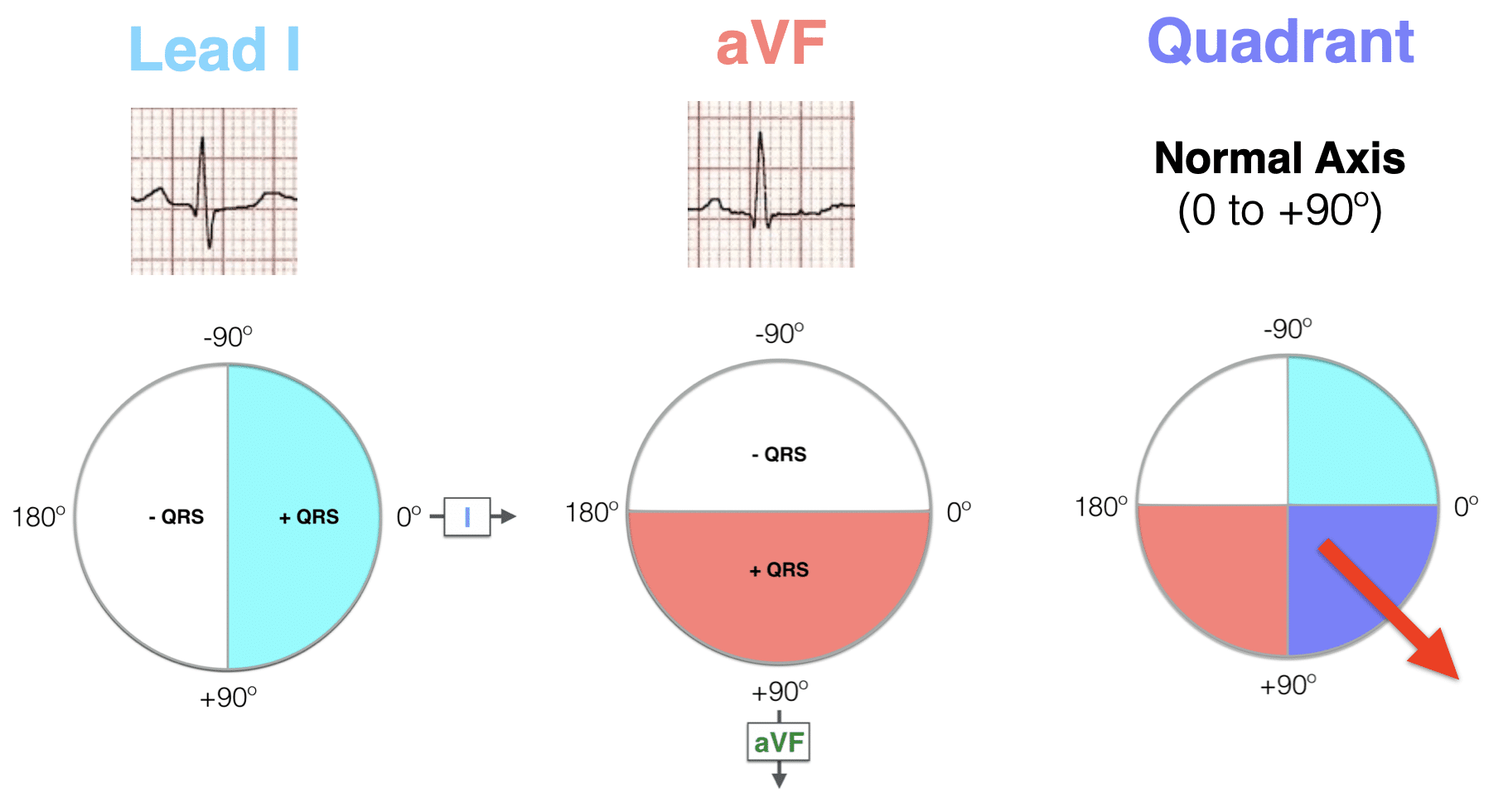
ECG Axis Interpretation LITFL ECG Library Basics
https://litfl.com/wp-content/uploads/2018/08/Hexaxial-ECG-AXIS-Quadrant-Method-Normal-axis-2021.png
https://ecgwaves.com/topic/ecg-normal-p-wa…
The P wave PR interval and PR segment ECG interpretation traditionally starts with an assessment of the P wave The P wave reflects atrial depolarization activation The PR interval is the distance between the

https://emedicine.medscape.com/article/2172196-overview
PR interval 120 200 milliseconds PR segment 50 120 milliseconds QRS complex 80 100 milliseconds ST segment 80 120 milliseconds T wave 160

Pin On PA Life

ECG Basics R E B E L EM Emergency Medicine Blog

Pin On Cardiology
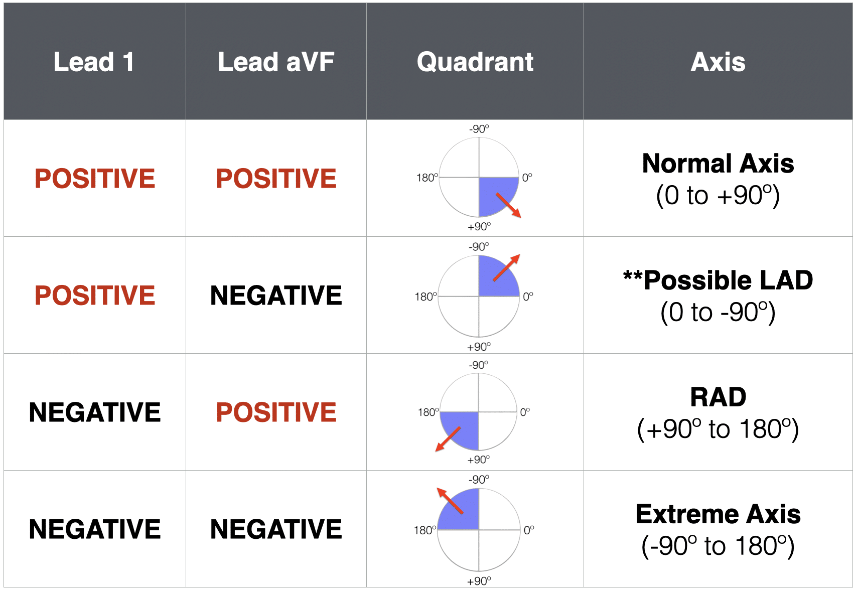
ECG Axis Interpretation LITFL ECG Library Basics

ECG Axis Interpretation LITFL ECG Library Basics
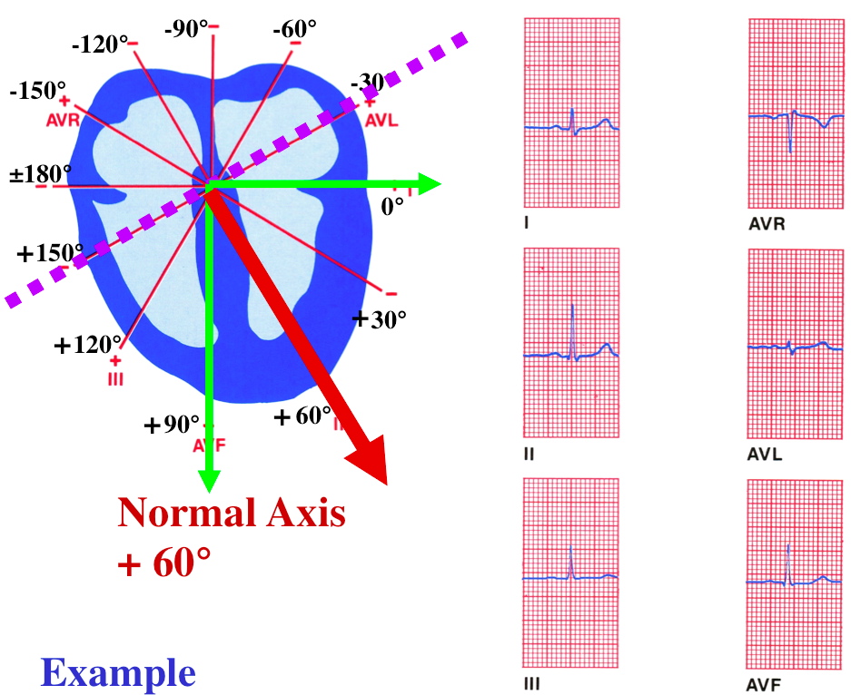
QRS Determination

QRS Determination
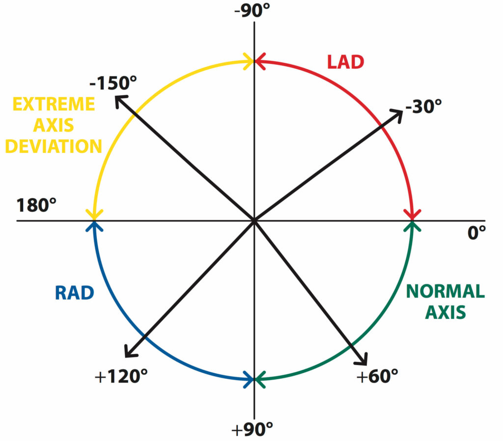
The Basics Of ECG Interpretation Part 2 Rate Rhythm And Axis
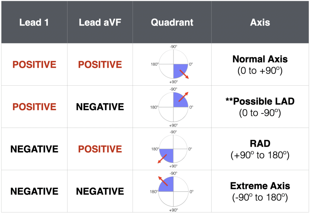
ECG Axis Interpretation LITFL ECG Library Basics
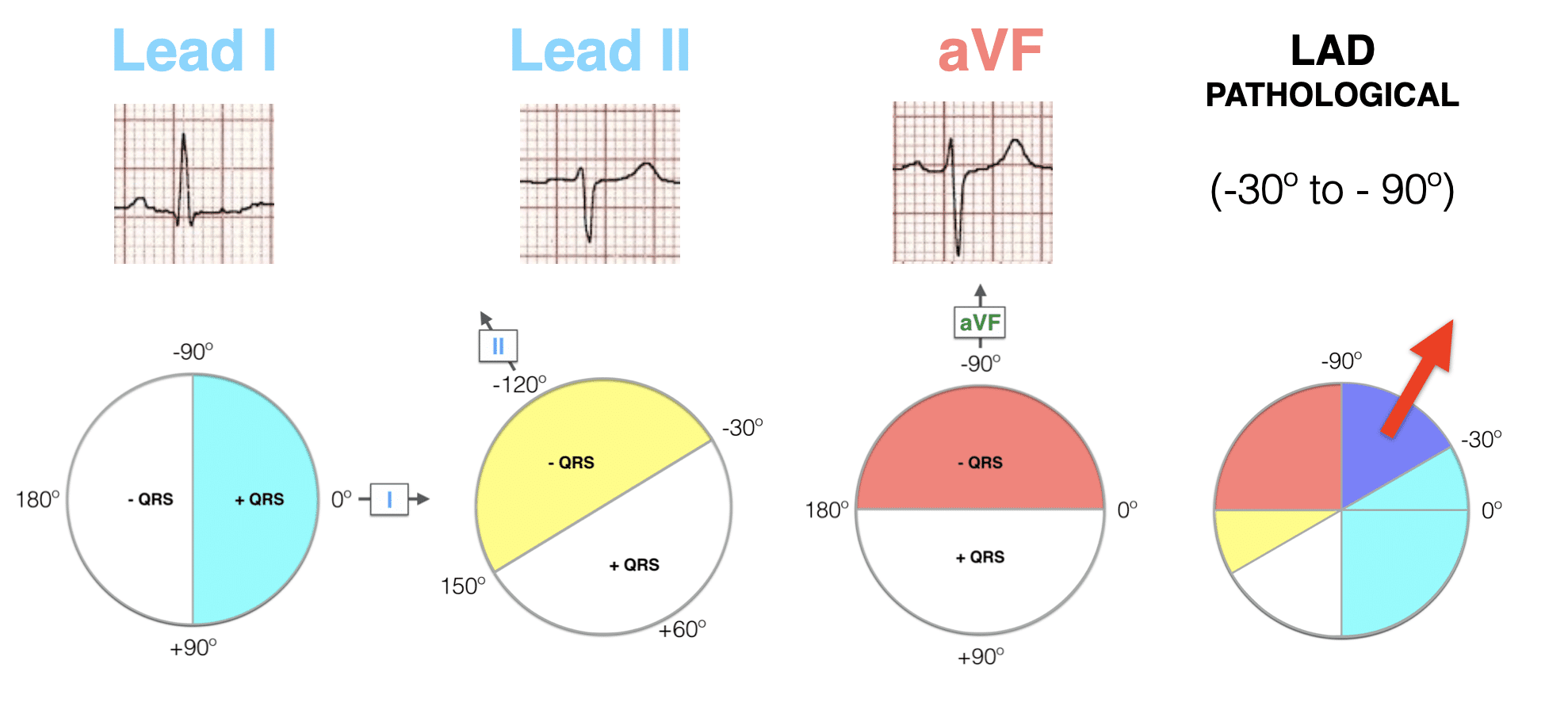
Left Axis Deviation LAD LITFL ECG Library Diagnosis
Ecg Frontal Axis P Normal Range - The mean frontal plane electrical QRS axis electrical axis usually points to the left and interiorly in the normal heart because of left ventricular dominance ECG