P R T Axes In Ecg Report Most ECG machines have in built interpreters that give all the P QRS and T axis Documented characteristics of a P wave mostly emphasis morphology duration and
The most efficient way to estimate axis is to look at LEAD I and LEAD aVF Examine the QRS complex in each lead and determine if it is Positive Isoelectric Right axis deviation Normal in newborns Right ventricular hypertrophy Acute cor pulmonale pulmonary embolism Chronic cor pulmonale COPD pulmonary
P R T Axes In Ecg Report

P R T Axes In Ecg Report
https://i.pinimg.com/originals/a5/47/6f/a5476f135062532b753565b8158e1cff.jpg

PDF 12 Lead ECG Wichita edu P R T Axes 61 52 97 Abnormal ECG When
https://img.pdfslide.net/doc/image/5ec8d6b120162707222fa0a7/12-lead-ecg-p-r-t-axes-61-52-97-abnormal-ecg-when-compared-with-ecg-of-17-may-2006.jpg
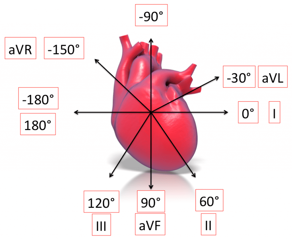
ECG Basics R E B E L EM Emergency Medicine Blog
http://rebelem.com/wp-content/uploads/2013/11/Axis-1024x839.png
As evident from the figure the normal heart axis is between 30 and 90 If the axis is more positive than 90 it is referred to as right axis deviation How to read an ECG There are many different systems to interpret the ECG This system ensures you will never miss anything Patient details Situation details Rate Rhthm Axis P wave and P R interval Q wave
During T P P R and S T segments electric field is stable and only subtle changes can be detected by skin electrodes These small changes of electric fields QRS axis QRS morphology P wave morphology PR interval QT interval ST segments T waves Any other deflections or waveforms in the ECG 5 2 Implicit and
Download P R T Axes In Ecg Report
More picture related to P R T Axes In Ecg Report
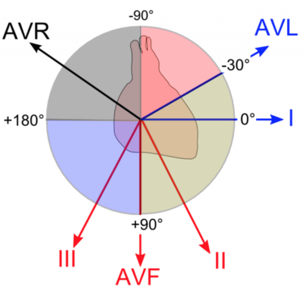
ECG Basics R E B E L EM Emergency Medicine Blog
http://rebelem.com/wp-content/uploads/2013/11/Axis-1-1024x976.png?a68ec4
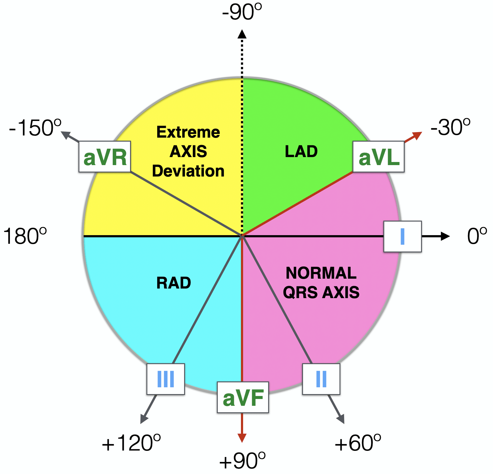
ECG Axis Interpretation LITFL ECG Library Basics
https://litfl.com/wp-content/uploads/2018/08/Haxaxial-ECG-AXIS-Reference-2021.png
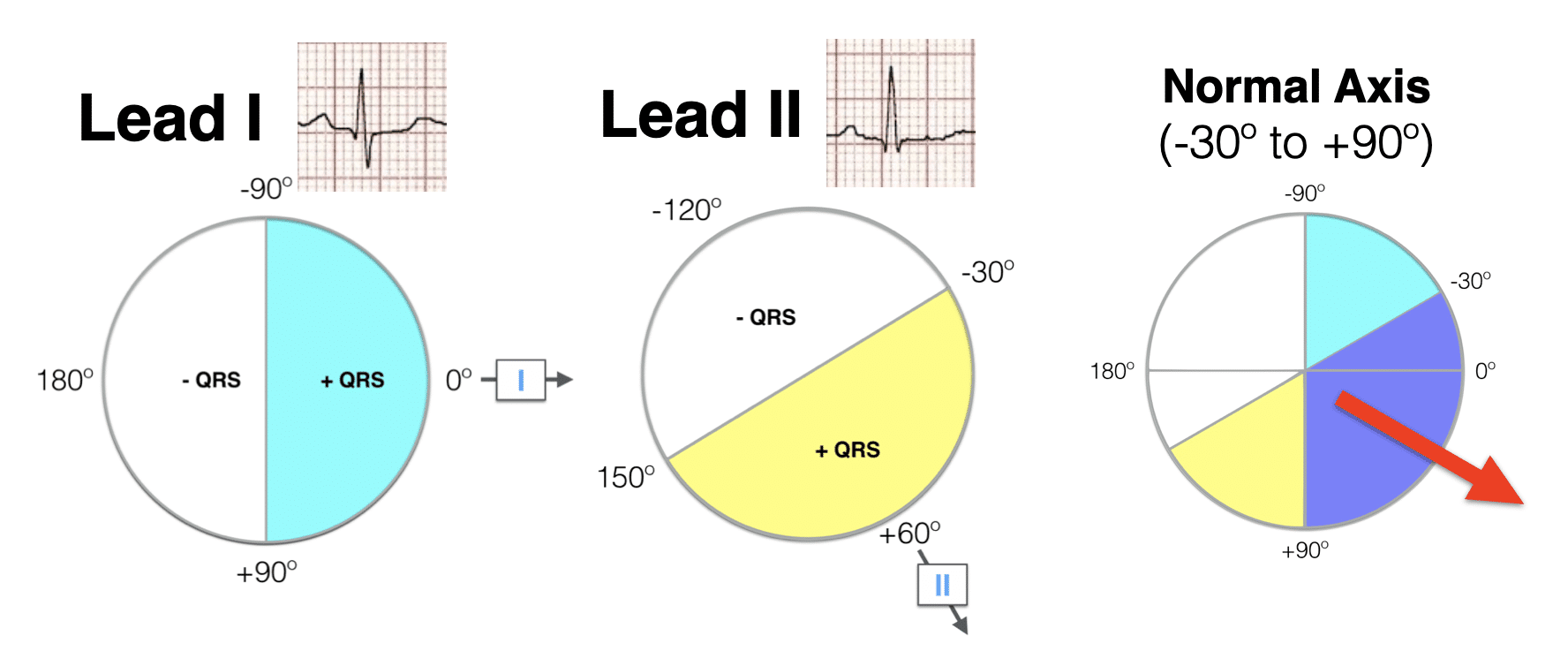
ECG Axis Interpretation LITFL ECG Library Basics
https://litfl.com/wp-content/uploads/2018/08/Lead-I-Lead-II-hexaxial-analysis-Normal-axis-2021.png
Normal ECG values for waves and intervals are as follows RR interval 0 6 1 2 seconds P wave 80 milliseconds PR interval 120 200 milliseconds PR segment Determine the heart rate by counting the number of large squares present on the EKG within one R R interval and dividing by 300 Identify the axis Know abnormal
Normative standards were developed for all 102 ECG variables including heart rate P R and T axis R T axis deviation PR interval QRS duration QT and QTc interval P Q R S and T amplitudes in 12 leads P Wave It is important to remember that the P wave represents the sequential activation of the right and left atria and it is common to see notched or biphasic P waves of right and

Pin By Holly Mitchell Arrington On Bacheca Frank Ecg Interpretation
https://i.pinimg.com/originals/fe/ec/46/feec469ab5fcc72cd86377b1d5b784c2.jpg

Ecg Interpretation Criteria Review Axis Deviation Left Right Hot Sex
https://slidetodoc.com/presentation_image/10ddc6347d6dd907f20b5875066172bb/image-2.jpg

https://irispublishers.com/ojcrr/fulltext/normal-p...
Most ECG machines have in built interpreters that give all the P QRS and T axis Documented characteristics of a P wave mostly emphasis morphology duration and

https://litfl.com/ecg-ax
The most efficient way to estimate axis is to look at LEAD I and LEAD aVF Examine the QRS complex in each lead and determine if it is Positive Isoelectric

Acute Myocardial Injury After Administration Of Intravenous Epinephrine

Pin By Holly Mitchell Arrington On Bacheca Frank Ecg Interpretation
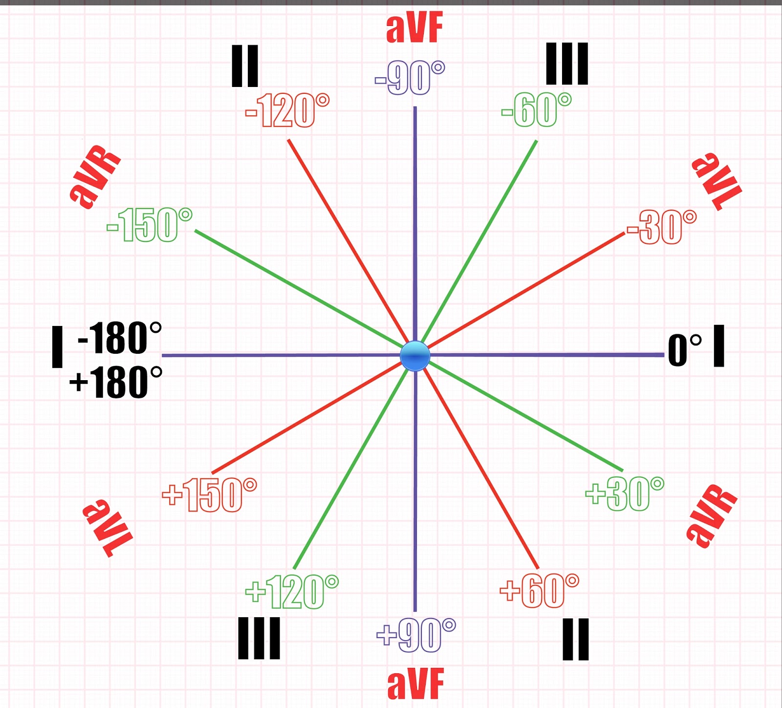
ECG Educator Blog Cardiac Axis Made Easy
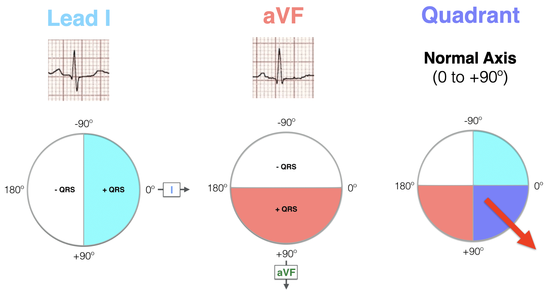
ECG Axis Interpretation LITFL ECG Library Basics
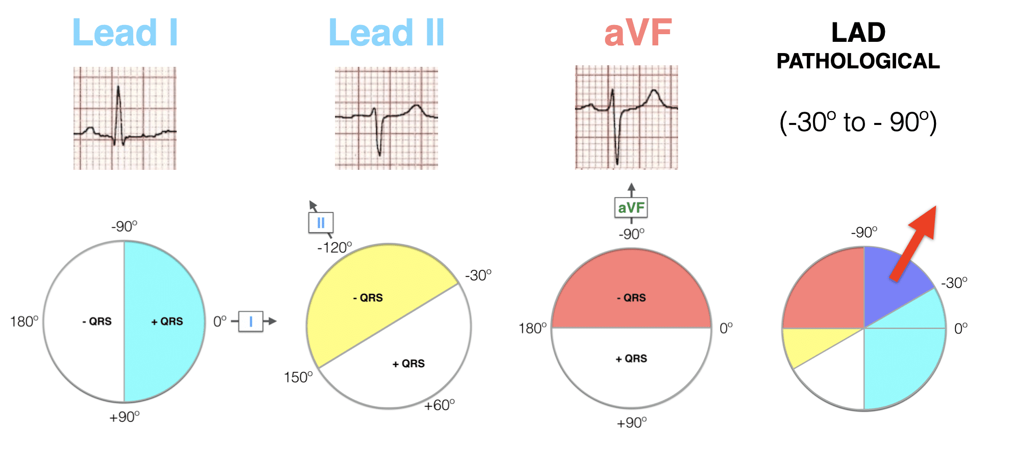
Left Axis Deviation LAD LITFL ECG Library Diagnosis
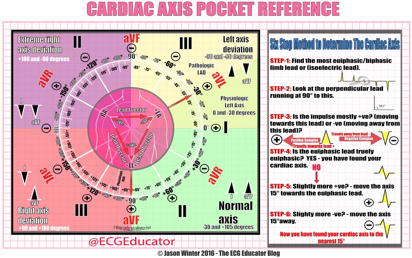
ECG Educator Blog Cardiac Axis Made Easy

ECG Educator Blog Cardiac Axis Made Easy
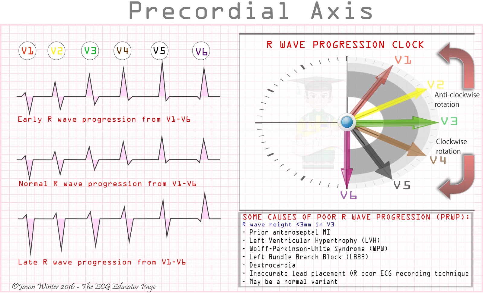
R Wave Progression Normal Chest Lead ECG Shows An GrepMed
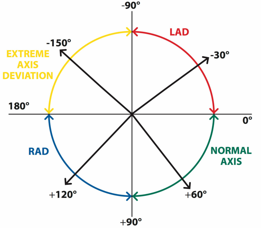
The Basics Of ECG Interpretation Part 2 Rate Rhythm And Axis

P Wave QRS Complex And T Wave Cardiac Physiology Nursing Study
P R T Axes In Ecg Report - During T P P R and S T segments electric field is stable and only subtle changes can be detected by skin electrodes These small changes of electric fields